• On-screen Digitising
  • Cephalometric Analysis
  • Surgical Prediction
  • Predictive Photo Morphing
  • Convenient Data Storage

OPAL is a powerful suite of programs for easy storage, sorting and manipulation of all clinical images - photographs, radiographs, study casts and any text documents. On-screen digitising of cephalometric x-rays and profile photographs, image enhancement and predictive profile morphing are all easy. Multiple cephalometric analyses, tracing superimpositions and treatment predictions are included.

OPAL is self-funding. Surplus funds go to the BOS foundation to support research and teaching.

OPAL is available at the BOS website at: www.bos.org.uk/Opal-Image-Sales.

COGSoft OPAL 2.5

September 10th, 2022

OPAL has undergone an extensive overhaul and upgrade to produce version 2.5. The principal new features include:

  • Option to digitise the 'stable' anatomical cephalometric features, enabling easy best-fit superimposition of tracings on these structures as is often required for case presentation.
  • Creation of fully customisable cephalometric analyses for research or clinical purposes on cephalometric radiographs or any other image e.g. a Little's index on a study model image.
  • Major internal overhaul to provide excellent compatibility with latest versions of Windows.
  • Longer file name suffixes enabling easier identification of different superimpositions.
  • Customisable and 'sticky' colours for each category of tracing e.g. for Start, Prediction, End of active treatment etc.
  • Improved morphing of the lips in prediction images
  • Removal of obsolete features e.g. digitising via an external digitiser and digitising of outlines by the individual entry of points.
  • Extensive updating and enhancement of the Help within OPAL

COGSoft OPAL 2.4

February 26th, 2014

New features in OPAL 2.4 include:

In OPAL Image Viewer

  • OPAL Image Viewer now creates tracings directly - no need to open the OPAL Tracing application. When you save a digitised cephalometric x-ray that has all required points and outlines digitised, a tracing will automatically also be saved. The resulting tracing will be tagged with the same date as the x-ray image and the same description marker if present (e.g. "(Start)"). If you subsequently edit the image again and save it, any corresponding tracing will be regenerated and overwritten.
  • The tendency for the lower lip to be prominent in predictions of treatment change has been addressed producing a more realistic prediction tracing or morphed profile image of the lower lip and reducing the need for any manual improvement of the prediction.
  • Context-sensitive scaling. When digitising on-screen (almost all current users) the scaling (magnification factor) must be set to 'fixed scale points' as described in OPAL Tracing. In OPAL 2.4, you will then be automatically prompted to enter these scale points when digitising an xray or study casts, but not when digitising a photographic profile. The rationale is that ceph xrays need scaling to give a meaningful ceph analyis. Profile digitising on the other hand is only done to enable best fit of that photo profile to an already digitised xray tracing soft tissue outline. When digitising study models it is assumed that you wish to make a millimetrically accurate analysis and that a scaling object e.g. a ruler has therefore been included in the image of the model to be digitised.

In OPAL Tracing

  • [ ] Plan Notes:
    This new function enables the entry of free text onto the prediction log. This enables important details such as the planned extractions to be recorded in the log and on the tracing.
  • It is now possible to save and export superimposed tracings. This works for both OPAL tracing files (OXT) and SVG files. Previously, superimpositions could be viewed and printed but not saved.
  • SVG tracings now support line styles - dashed and dotted lines.

In OPAL Explorer

  • OPAL Explorer supports pasting of 32 bit images from the clipboard in addition to 24 bit ones. For example, this means that if your monitor is set for 32 bit colour then screenshots copied to the Windows clipboard will be pastable straight into OPAL. In addition to the general usefulness of this ability to save good quality screenshots in OPAL, this function has for example enabled one user whose PACS would not export jpeg images to easily export xrays to OPAL.
  • Fixed bug that was causing "Open with..." to not work on Windows 7 and above to display.

Videos

July 12th, 2010

The Screenshots page now includes some videos of OPAL 2.3 in action.

Play

COGSoft OPAL 2.3

February 12th, 2010

New features in OPAL 2.3 include:

In OPAL Explorer

  • Search the patient database rapidly by directory or by entire database
  • 9 - photo montage to include 3/4 face view
  • More rapid re-orientation of images - especially 'mirror' photos
  • Ini file mapping for storing of personal user preferences
In OPAL Image Viewer
  • Rapid, smooth digitising of outlines using Bezier curves
  • Enhanced undo/redo permits temporary enhancement of all or part of an image whilst digitising
  • Audio prompts for on-screen digitising point entry
  • Option for entry of just a few points gives rapid Eastman analysis
  • Improved, smoother morphing of lip profiles
  • Enhanced wizard for saving morphed images in prediction montages
  • On-screen measurement of irregular areas in square millimetres or pixels
In OPAL Tracing (formerly called OPAL xray)
  • Left-Right switch for profile direction.
  • Tracings can be saved as SVG files for smoother outlines, greatly enhanced editing and for viewing without OPAL - useful for presentations or for e-mailing tracings.
  • Recalibrated Bolton Standards give better shape comparisons.
In OPAL HELP
  • Word-searchable HELP - find the required HELP more easily

Inkscape

February 12th, 2010

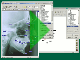
OPAL can save x-ray tracings as SVG files that can be edited using Inkscape. In Inkscape you can modify line thicknesses, change fonts, remove unwanted parts of the tracing, add captions, combine with other images - the list is endless. Inkscape can then export modified tracings as bitmap images which can be imported into Powerpoint, etc.

Inkscape is an Open Source vector graphics editor, with capabilities similar to Illustrator, CorelDraw, or Xara X, using the W3C standard Scalable Vector Graphics (SVG) file format.

The outline editing using Bezier curves first introduced in OPAL 2.3 is designed to work in a similar way to the curve editing provided by Inkscape - so once you are comfortable with curve editing in either program, you will probably find it easy to edit curves in the other.

OPAL Version 2.2

June 21st, 2007

OPAL 2.2 updated major new functions which were introduced in version 2.1.

The changes for 2.2 from 2.1 focussed on:

  • a need to cope better with the increasingly common situation where a computer does not have a COM port.
  • a need to address a potential scaling problem when dumping previously-digitised coordinates to create a new tracing.
  • a need to update the HELP in some areas.

OpalImage Version 2.1

September 22nd, 2005

  • IMAGE MORPHING.
    A photograph of a patient's profile can be automatically 'morphed' in step with changes to a corresponding overlaid cephalometric tracing. As a consequence, it is possible to give an idea as to how the patient's appearance will change as a result of surgical procedures.
  • PREDICTION MONTAGE.
    A new type of montage displaying just two photographs side by side. Typically, the left hand image will be an original profile photograph and the right hand image will be a morphed prediction image based on the original profile.
  • IMAGE AND TRACING SAVE HELPER.
    This feature assists with the saving of morphed images in a prediction montage and the corresponding prediction tracing - attempting to give them both the same new corresponding label and short description.
  • DATASET RECOMMENDATIONS AND OUTCOME AUDIT FOR ORTHOGNATHIC PATIENTS.
    This is provided through the generation of standard documents, based on templates, filling in certain fields automatically. As distributed, two template documents are provided, namely 'Surgical Orthodontic Analysis' and 'Surgical Orthodontic Minimum Dataset'. The first of these enables easy auditing of the cephalometric outcome of orthognathic treatment and the second assists the full collection of the data recommended by BOS and BAOMS for these patients.

OpalImage Version 2.0

December 9th, 2003

OpalImage 2.0 incorporated major new functions over previous versions.

  • A COMPLETELY NEW FILE STRUCTURE
    Although largely 'invisible', this is a powerful tool for increased robustness and flexibility.
  • ON-SCREEN DIGITISING
    OpalImage 2.0 supports digitising cephalometric radiographs on-screen. Digitising for clinical purposes does not require a (potentially expensive) digitising tablet.
  • SUPERIMPOSITION OF A TRACING ON A PATIENT PROFILE PHOTOGRAPH
    A cephalometric tracing can be overlaid on a photograph of the profile of a patient by dragging and dropping. The tracing is automatically aligned using selected common reference points.
  • IMAGE ENHANCEMENT within OPAL-Image.
    The Opal image viewer can adjust contrast, brightness, hue, colour and intensity curves on the whole image or any selected region. This can be done even in the middle of on-screen digitising to help make points and outlines clearer.
  • ARTISTIC MODIFICATION OF OUTLINES.
    Outlines can be adjusted by hand using various tools to better represent the probable current or predicted profile.
  • SEARCH FUNCTION.
    The database has an easy and capable search capacity for finding patients or individual files within patient folders with chosen features.
  • DATES IN NEW INTERNATIONAL STANDARD FORMAT (ISO-8601).
    Although less familiar in every-day use in the U.K., this avoids international confusions and permits more logical sorting of files in date order using any program.